Evaluation of the Fetal T/R Ratio Using a Fetal
Scalp Electrode and Abdominal Sensors

Caitlin McDonnell 1 ; Reza Sameni, PhD 2,3 ; Jay Ward 3 ; Jim Robertson 3 ;
Adam Wolfberg, MD, MPH 3,4 ; Gari Clifford, PhD,1,3
1University of Oxford, 2Shiraz University, 3MindChild Medical, Inc.,
4Tufts Medical Center
Results:
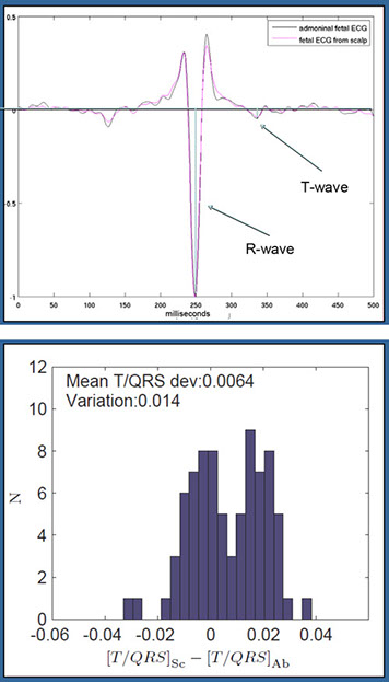
The difference between the T/R ratio calculated from the scalp electrode and the T/R ratio calculated from the extracted abdominal fetal ECG was 0.0064 ± 0.014. This difference is not clinically meaningful.
Conclusion:
We measured the fetal T/R ratio was accurately measured using abdominal electrodes in non-ischemic fetuses.
Fig. 1
Comparison of ECG waveform from
the FSE and abdominal sensors.
The T‐ and R‐waves are illustrated.
Fig. 2
Variation in T/R ratio between
FSE and abdominal sensors.
Background:
Evaluation of the T/R ratio - the
metric used by the STAN™
monitor - improves the accuracy of intra-partum fetal assessment when combined with fetal heart rate monitoring, but typically requires a fetal scalp electrode (FSE). Noninvasive measurement of the T/R ratio would make this metric more widely available.
Objective:
To compare fetal T/R ratio measured using sensors on the maternal abdomen to the fetal T/R ratio acquired using a FSE.
Methods:
Data were acquired from 27 term laboring women who had a FSE placed for a clinical indication. 31 channels of abdominal data were recorded simultaneously with the FSE.
The average T/R ratio level was estimated from 79 30-second segments from the FSE and the abdominal data for 4 subjects. A comparison was performed to assess the correlation between the fetal T/R ratio derived from abdominal sensors and T/R ratio measured using the FSE.
Disclosure: The authors, except for Ms. Pettigrew, hold equity in MindChild Medical, Inc., which has licensed intellectual property used to generate results presented in this abstract.

© 2021-2022 Mindchild Medical, Inc. | 1600 Osgood Street | Suite 2017 | North Andover, MA 01845 | info@mindchild.com | 978.566.9880